- C-MORE Home
- What is Microbial Oceanography?
- What is C-MORE?
- Research
- • Research Cruises
- » BioLINCS
- Home
- Cruise Participants
- Instruments Used
on the Cruise - Nitrogen Cycling
- Marine Microbes
- Cruise Blog
- Data Archive
- Education & Outreach
- People
- Publications
- Image Library
- Contact Us
| |||||||||||||||||||||||||||||||||||
BioLINCS Cruise Blog
Monday, September 12, 2011
|
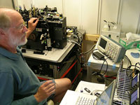
Ger van den Engh adjusts the lasers on his influx flow cytometer (IFC) on board the Kilo Moana. Ger invented this amazing machine, and was excited to see how useful it was for the microbiologists on board the cruise. |
|
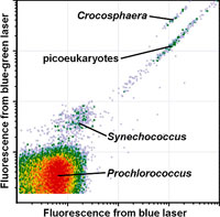
This graph created by the influx flow cytometer (IFC) shows microbes in a sample of seawater from 45 meters below the surface. The location of each dot indicates how much each individual microbe glowed (fluoresced) as it passed in front of light beams from a blue laser and a blue-green laser. Each “cloud” of dots represents a different type of microbe with different pigments. |
|
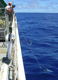
John Waterbury pulls a plankton net up through the water off the stern of the Kilo Moana. The water is so clear that the net looks as though it is floating in air. But it nonetheless harbors an abundance of microscopic organisms. |
|
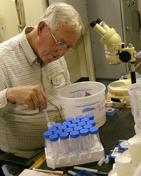
John Waterbury sorts through the plankton that he collected by hand using a plankton net. Using a microscope and a pipette, he picked out different types of Trichodesmium bacteria, which he will try to culture in his lab back at Woods Hole Oceanographic Institute. |
Yesterday I talked a little bit about how we can identify different marine bacteria using “molecular biology” techniques and DNA analyses. In this blog, I’ll focus on an amazing instrument we have on board the ship called the influx flow cytometer (IFC). The IFC can help identify microbes based on their size and on the types of pigments they carry inside their cells.
Most influx flow cytometers are purchased by biomedical research groups, which use them to sort blood cells (which contain the pigment hemoglobin) or transparent cells that have been treated with fluorescent dyes. However, photosynthetic bacteria and algae make great subjects for the IFC because they naturally contain a variety of pigments that they use in the process of extracting energy from sunlight.
Ken Doggett, of the University of Hawai‘i, has been using this instrument to study marine algae and bacteria for several years. For this cruise, he invited the inventor of the instrument, Ger van den Engh, to come along and see how the instrument is being used for cutting-edge marine microbiology research. Ger recently gave me a ”tour” of the IFC that left me shaking my head in wonder.
At the core of the instrument is a tiny jet of water, about the diameter of a human hair, which passes in front of four laser beams of different colors—yellow-green, turquoise, blue-green, and purple. Individual bacteria or cells can be injected into the exact center of this jet of water so that the cells pass in front of each of the laser beams in single file, one at a time. When the light from the lasers hits pigments, such as chlorophyll, inside the cells, they make the pigments glow (fluoresce). Sensitive light detectors inside the IFC measure the fleeting glimmer of light given off by each individual cell as it passes through the machine.
Different types of microbes have different amounts and types of pigments inside their cells. By comparing the amount of fluorescence excited by each of the four lasers, researchers can tell one type of microbe from another, as they pass through the instrument. This alone is amazing, considering that some marine bacteria are about 1/100th the diameter of a human hair, and that they pass in front of the lasers at about 60 miles an hour.
In the graph at right, you can see how the IFC can provide an indication of what kinds of marine microbes are present in a given water sample. This graph shows the relative fluorescence of microbes that were exposed to two of the four lasers in the IFC (the blue laser and the blue-green laser). Each dot on this graph represents a single microbe passing through the lasers. Each ”cloud” of dots represents a different group of organisms.
At the lower left of the graph is a big, dense ”cloud” which shows a whole lot of Prochlorococcus bacteria. As I noted in yesterday’s blog, these are the dominant photosynthetic microbes in the open ocean waters around the world, and account for the ”deep chlorophyll maximum” that we see in our CTD casts.
Just above and to the right of the Prochlorococcus ”cloud” is a smaller cloud of points that represent individual cells of another marine bacterium, Synechococcus. Synechococcus is probably the second most abundant type of photosynthetic marine bacterium in open ocean areas, after Prochlorococcus.
Near the top of the graph is a small line of points that represent cells of Crocosphaera, a somewhat larger type of marine bacterium. Unlike Prochlorococcus and Synechococcus, Crocosphaera is a ”nitrogen-fixing” bacterium, and is only found in waters above 24° Celsius (75° Fahrenheit). But you can’t tell that from looking at this graph.
Finally, the long line of dots extending to the upper right of the graph are believed to represent cells of a diverse group of photosynthetic algae called “picoeukaryotes.” These are tiny algae, but they are still larger than many marine bacteria.
Learning to identify different types of marine microbes based on these graphs takes a lot of time and experience with the IFC—two things that Ken Doggett has quite a bit of. It also involves taking organisms out of the cytometer and identifying them using a microscope or DNA analysis.
This brings up another amazing aspect of the IFC—it can pick out individual bacteria, one at a time, from the stream of water, and place them into different vials, based on their size and fluorescence. It does this by using ultrasonic sound to to break up the stream of water into thousands of microscopic drops, each of which holds a single bacterial cell. Then it uses an electrostatic field (like the charge generated by static electricity) to pull specific drops of water out of the main stream and aim these drops so that they land in a vial at the bottom of the instrument.
For example, if Ken wants to study Crocosphaera bacteria, he tells the IFC to look for cells that are the size and fluorescence of these bacteria. If a cell matches the selected criteria, the IFC directs the microdrop containing that cell into a specific vial. After a while, Ken has a tiny vial containing a tiny bit of seawater and hundreds or thousands of Crocosphaera cells. He can then do DNA analysis on those cells, knowing that they’re not mixed up with the cells of a bunch of other types of microbes.
After watching Ger’s amazing science-fiction machine for a while, I walked out to the back deck of the Kilo Moana. There I saw John Waterbury applying a sampling technique that marine biologists have used for well over a century—pulling a conical net of sheer white fabric through the water, over and over again.
After five or ten passes, John finally brought the ”plankton net” up on deck and then washed the contents into a white plastic bucket. The bucket was full of tiny swimming creatures (copepods), as well as lots of little brown hairs and ”puff balls” about a millimeter or two across.
The hairs and puff balls were our old friend Trichodesmium, which I described in my September 7 blog entry. After looking through his catch using a microscope, John picked out individual Trichodesmium colonies and carefully set them aside, with the hope of growing them in his laboratory back in Woods Hole. He suspects that the hairs and puff balls are actually different strains of the bacteria.
John is an expert on the culture of marine bacteria, which are notoriously difficult to grow in the lab. For decades he has applied basic science, careful experimentation, and a huge dose of patience to develop techniques for growing microbes that nobody else could grow.
It was fascinating to discover that there is still room in modern research for both the influx flow cytometer and the hand-pulled plankton tow. Personally, I find equal inspiration in a machine that can sort individual bacteria, one at a time, and a bucket full of wriggling creatures and pale brown slime…
[ Top of Page ]


